BLACK FRIDAY DEALS

  • Home
  • About
  • Product
  • Patient Corner
  • Doctor's Corner
  • Testimonial
  • FAQs
  • Contact
  • More
    • Home
    • About
    • Product
    • Patient Corner
    • Doctor's Corner
    • Testimonial
    • FAQs
    • Contact
  • Sign In
  • Create Account

  • Orders
  • My Account
  • Signed in as:

  • filler@godaddy.com


  • Orders
  • My Account
  • Sign out

Signed in as:

filler@godaddy.com

  • Home
  • About
  • Product
  • Patient Corner
  • Doctor's Corner
  • Testimonial
  • FAQs
  • Contact

Account


  • Orders
  • My Account
  • Sign out


  • Sign In
  • Orders
  • My Account

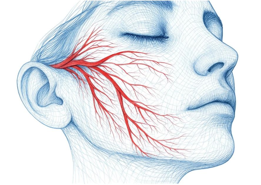
Vasodilation

Vasodilation means your blood vessels widen, allowing more blood to flow to the skin. This helps deliver more oxygen and nutrients to skin cells, which can support collagen production and keep your skin healthy. While vasodilation does not create collagen, it creates the ideal environment to repair and renew itself.

How to assess, educate, and treat various skin conditions

1. Basic Clinical Observation

  • Small, flat, light-to-dark brown spots, most visible on sun-exposed areas.
  • Typically become more pronounced after sun exposure and may fade during winter months.

2. Contributing Factors

  • Genetics: Frequently observed in individuals with lighter skin types.
  • Sun exposure: UV radiation increases the visibility of freckles.
  • Skin type: Fair skin with less natural melanin protection is more prone.
  • Aging: With age, freckles may fade, while lentigines (age spots) become more common.

3. Commonly Referenced Tools

  • Clinical exam: Visual inspection under natural light.
  • Wood’s Lamp: Assists in differentiating freckles from deeper pigmentation or lentigines.
  • Dermatoscope: Provides magnification to help distinguish freckles from other pigmented lesions.

4. Clinical & Preventive Considerations

  • Prevention: Daily use of broad-spectrum sunscreen, minimizing UV exposure, and protective clothing.
  • Management references: Brightening skincare ingredients such as vitamin C and niacinamide; professional options like peels or light/laser procedures may be considered under physician guidance.
  • Supportive care: Thermal-based devices (e.g., VASO Mask) may be incorporated as part of skincare routines to support circulation and help maintain even skin appearance.


1. Basic Clinical Observation

  • Visual Inspection:
    Telangiectasia refers to small, visible dilated blood vessels near the skin surface. They are most commonly found on the face (nose, cheeks, chin) or lower extremities.
  • Common Patterns:
    • Linear: Straight or slightly curved red lines.
    • Arborizing: Branching, tree-like appearance.
    • Punctate: Small, dot-like red spots.
    • Matting: Fine clusters of vessels that resemble spider veins.

2. Contributing Factors

  • Genetic tendency: Family history of vascular fragility.
  • Sun exposure: Long-term UV damage can contribute.
    Underlying skin conditions: Rosacea, chronic dermatitis.
  • Hormonal influences: Pregnancy, contraceptive use, or hormonal shifts.
  • Lifestyle / environmental factors: Alcohol consumption, smoking, extreme heat or cold exposure.
  • Aging: Natural increase in vascular fragility with age.

3. Commonly Referenced Assessment Tools

  • Dermatoscope: Helps visualize vessel morphology in detail.
  • High-resolution or 3D imaging: Used to document severity and monitor changes over time.
  • Clinical photography: Commonly used for before-and-after comparisons.

4. Clinical & Preventive Considerations

  • Prevention (general measures):
    Sun protection, gentle skincare routines, avoiding excessive heat or cold, and minimizing lifestyle triggers such as alcohol or smoking.
  • Management Options (commonly referenced in dermatology):
    • Topical care: Anti-inflammatory or barrier-repair formulations to support skin health and reduce visible redness.
    • Energy-based devices: Vascular lasers (e.g., pulsed dye laser, Nd:YAG) or IPL, generally considered under professional evaluation.
    • Adjunctive / supportive care: Thermal-based devices (e.g., VASO Mask) may support circulation and overall skin appearance, though typically positioned as complementary.


1. Basic Clinical Observation

    Visual Inspection:Enlarged pores appear as small openings on the skin surface, most    

    noticeable on the nose, cheeks, and forehead.

     Characteristics:

  • Size may vary depending on skin type and sebum activity.
  • More prominent in oily or combination skin.
  • Can appear as round or oval openings.

2. Contributing Factors

  • Genetics: Some individuals naturally have larger pores.
  • Sebum production: Higher oil secretion can make pores more visible.
  • Skin aging: Loss of elasticity and collagen may make pores appear larger.
  • Sun exposure: UV damage weakens skin support structures, contributing to pore visibility.
  • Lifestyle: Poor cleansing habits, smoking, and environmental pollution may influence pore appearance.

3. Commonly Referenced Tools

  • Clinical examination: Direct visual assessment under natural or magnified light.
  • Dermatoscope / Magnification: Helps visualize pore size and distribution in more detail.
  • 3D skin imaging: Provides quantitative analysis of pore density and size for documentation and follow-up.

4. Clinical & Preventive Considerations

     Prevention (general measures):

  • Consistent daily sunscreen use to protect collagen from UV damage.
  • Regular gentle cleansing to reduce oil and debris buildup.

     Management (commonly referenced in dermatology):

  • Topical care: Retinoids (e.g., retinol), niacinamide, and mild exfoliating agents are often included in skincare routines to support smoother skin appearance.
  • Professional options: Chemical peels, laser, or energy-based devices may be considered under physician guidance for patients seeking improvement.

     Supportive care: Thermal-based devices (e.g., VASO Mask) may be incorporated as adjunctive      

    care to support circulation and overall skin condition.


1. Basic Clinical Observation

    Visual Inspection:Assess the distribution, intensity, and pattern of dyspigmentation (e.g.,    

   freckles, melasma, sunspots, age spots).

    Classification (commonly referenced in dermatology):

  • Epidermal dyspigmentation: Light to dark brown, often more responsive to topical agents or light-based therapies.
  • Dermal dyspigmentation: Deeper, bluish-gray tone, generally more resistant to treatment.
  • Mixed type: Combination of both epidermal and dermal dyspigmentation.

     Pattern & Localization:

  • Diffuse: Generalized darkening
  • Localized: Spots or patches
  • Symmetrical vs. Asymmetrical: e.g., melasma vs. post-inflammatory dyspigmentation.

2. Factors Contributing to Dyspigmentation

  • Sun Exposure: UV radiation is a primary trigger.
  • Hormonal Influences: Pregnancy, oral contraceptives may contribute to melasma.
  • Post-Inflammatory Dyspigmentation (PID): Occurs after acne, eczema, or skin injury.
  • Genetics & Skin Type: Darker skin types (higher Fitzpatrick types) are more prone to visible dyspigmentation.
  • Aging: May lead to solar lentigines (age spots) and uneven skin tone.

3. Professional Assessment Tools

  • Wood’s Lamp Examination: Helps differentiate epidermal vs. dermal dyspigmentation.
  • Dermatoscope: Provides magnified visualization of dyspigmentation patterns.
  • 3D Imaging / Spectrophotometry: Quantifies dyspigmentation intensity and monitors treatment progress.

4. Clinical Recommendations

      Prevention (widely recommended):

  • Daily broad-spectrum sunscreen, UV avoidance, maintaining hydration.

      Management Options (commonly referenced in clinical practice):

  • Topical agents: Vitamin C, niacinamide.
  • Chemical peels: Superficial peels (e.g., AHA, TCA) for epidermal dyspigmentation.
  • Energy-based devices: Laser or IPL, under professional supervision.
  • Adjunctive / supportive care: Thermal-based devices (e.g., VASO Mask) may support circulation and contribute to a more even skin tone.


1. Basic Clinical Test

Pinch Test (Skin Turgor Test)
Method: Use your thumb and forefinger to lightly pinch the skin (commonly on the back of the hand or under the eye) for 5 seconds.

Interpretation:

  • 1–2 sec → Healthy elasticity (typical for individuals under 30)
  • 3–4 sec → Functional age 30–44
  • 5–9 sec → Functional age 45–50
  • 10–15 sec → Functional age ≈ 60
  • >15 sec → Severe elasticity loss, common in individuals over 60

2. Factors Affecting Elasticity

  • Intrinsic Aging: Natural decline of collagen and elastin with age.
  • Photoaging: UV exposure damages collagen and accelerates elasticity loss.
  • Smoking: Reduces skin oxygenation, contributing to premature aging.
  • Extreme Weight Loss: Causes loose, sagging skin.
  • Chronic Dehydration: Decreases skin firmness and turgor.

3. Professional Measurement Tools

  • Cutometer: Uses suction to measure skin deformation and recovery; widely applied in clinical studies of collagen and elasticity.
  • High-frequency Ultrasound: Evaluates dermal thickness and collagen density, indicating structural aging.
  • 3D Skin Imaging: Provides visual analysis of collagen and elastin fiber distribution, enabling before-and-after comparisons.

4. Clinical Recommendations

        Preventive Care:

  • Sun protection, hydration, balanced diet, smoking cessation.

       Therapeutic Options:

  • Thermal therapy: e.g., VASO Mask, RF, infrared
  • Energy-based devices: laser, RF, ultrasound
  • Injectables: hyaluronic acid, collagen stimulators

       Topical agents: vitamin C, peptides, antioxidants


1. Basic Clinical Observation

Visual Inspection: Skin texture refers to the smoothness and uniformity of the skin surface.

Common Characteristics:

  • Uneven texture may appear as roughness, bumps, or enlarged pores.
  • Smooth texture indicates healthy turnover and hydration.

2. Contributing Factors

  • Intrinsic aging: Natural slowing of cell turnover.
  • Sun exposure: UV damage leads to roughness and uneven tone.
  • Dehydration: Lack of hydration reduces smoothness.
  • Excess sebum / acne: Clogged pores and breakouts affect surface texture.
  • Lifestyle factors: Poor cleansing, smoking, and pollution worsen texture.

3. Commonly Referenced Tools

  • Clinical exam: Visual and tactile evaluation of roughness and smoothness.
  • Dermatoscope / magnification: Allows closer inspection of unevenness and pores.
  • 3D skin imaging: Quantifies surface irregularities and tracks improvement after treatments.

4. Clinical & Preventive Considerations

Prevention (general measures):

  • Daily sunscreen, proper hydration, gentle cleansing, balanced diet.

Management (commonly referenced in dermatology):

  • Topical care: Retinoids, niacinamide, and vitamin C are often included in routines to support smoother texture.
  • Professional care: Options such as chemical peels, microdermabrasion, or energy-based devices may be considered under physician evaluation.

Supportive care: Thermal-based devices (e.g., VASO Mask) may be incorporated to support circulation and overall skin health.


1. Basic Clinical Observation

  • Visual Inspection:
    Assess wrinkle location, depth, and length (e.g., forehead lines, crow’s feet, nasolabial folds).
  • Classification by Depth:
    • Fine lines: Superficial, often linked to dehydration or sun exposure.
    • Moderate wrinkles: Extend into the dermis, associated with collagen loss.
    • Deep wrinkles: Prominent folds, commonly around the mouth and eyes.
  • Dynamic vs. Static Wrinkles:
    • Dynamic wrinkles: Appear only with facial expressions (smiling, frowning).
    • Static wrinkles: Visible even when the face is at rest.

2. Factors Affecting Wrinkle Formation

  • Intrinsic Aging: Natural reduction of collagen and elastin.
  • Photoaging: UV exposure damages dermal structures.
  • Lifestyle: Smoking, poor diet, dehydration accelerate wrinkle development.
  • Facial Expressions: Repeated muscle activity leads to gradual fixation of lines.

3. Professional Measurement Tools

  • Glogau Photoaging Scale
    Ranges from I–IV, classifying wrinkles from mild to severe with aging changes.
  • 3D Skin Imaging
    Quantifies wrinkle depth, length, and distribution, useful for before–after comparisons.
  • Silicone Replica Method
    Creates a skin impression analyzed under microscopy to evaluate wrinkle morphology.

4. Clinical Recommendations

  • Preventive Care:
    Daily sun protection, hydration, antioxidant-rich diet, smoking cessation.
  • Therapeutic Options:
    • Topical agents: Retinoids, peptides, antioxidants.
    • Injectables: Botulinum toxin (for dynamic wrinkles), hyaluronic acid or collagen stimulators (for static/deep wrinkles).
    • Energy-based therapies: Laser, RF, ultrasound, thermal devices (e.g., VASO Mask).


Patient Education

Intrinsic Aging Factors - Uncontrollable

Intrinsic Aging Factors - Uncontrollable

Intrinsic Aging Factors - Uncontrollable

• Age – Wrinkling, lentigines, and telangiectasia increase with age. 

• Gender – Males show more wrinkling/telangiectasia; females influenced by 

menopause and hormones.

• Ethnicity – Asians show more dyspigmentation; Europeans show earlier wrinkling.

• Hormonal – Menopause affects skin aging, though findings remain mixed.

Extrinsic Aging Factors - Controllable

Intrinsic Aging Factors - Uncontrollable

Intrinsic Aging Factors - Uncontrollable

• Nutrition – Higher vegetable and fatty acid intake reduces wrinkles/dryness.

• Sleep – Good sleep quality linked to reduced skin aging.

• Sunscreen – Use reduces wrinkling and photoaging.

• Smoking – Strongly accelerates wrinkling (“smoker’s face”).

• Air Pollution – Linked to lentigines, wrinkles, sagging.

• Lifestyle – Stress and sleep deprivation cause wrinkles, droopy eyelids.

• Alcohol – High intake (>40 g/day) strongly linked to wrinkles.

Treatment Pathway

Stage 1

Stage 1

Stage 1

Discuss extrinsic and intrinsic aging factors

• Moisturizer / Hydration → e.g., hyaluronic acid, aloe vera, ceramides

• Sunscreen (SPF 30+) → broad-spectrum, daily use


  • IN OFFICE VASO MASK USE : Vaso Mask quarterly with post chemical adsorption utilizing doctors choice hydration, rejuvination chemical.
  • AT HOME TREATMENT : Vaso Mask as needed with post chemical adsorption utilizing doctors choice hydration, rejuvination chemical.


Stage 2

Stage 1

Stage 1

All of Stage 1 Plus (+ –)

• Antioxidants Vitamin C, Vitamin E, Niacinamide (B3)

• Aesthetic non-invasive procedures → e.g., chemical peels and Hydrafacial for gentle skin renewal


  • IN OFFICE VASO MASK USE: Vaso Mask monthly with post chemical adsorption utilizing doctors choice hydration, rejuvination chemical.
  • AT HOME VASO MASK USE:  Vaso Mask Every 2 weeks with post chemical adsorption utilizing doctors choice hydration, rejuvination chemica

Stage 3

Stage 3

Stage 3

All of Stage 1,2 treatment,plus

• Injectables and Dermal fillers

• Thermal energy that heats the dermis Intense pulsed light (IPL), Radiofrequency (RF) and Laser


  • IN OFFICE VASO MASK USE: Vaso Mask weekly with post chemical adsorption utilizing doctors choice hydration, rejuvination chemical.
  • AT HOME VASO MASK USE:  Vaso Mask weekly with post chemical adsorption utilizing doctors choice hydration, rejuvination chemical.

Stage 4

Stage 3

Stage 3

All of Stage 1,2,3 treatment,plus

• Plastic surgery→ (e.g., facelifts or eyelid surgery)

• Hormone replacement therapy (HRT)


  • IN OFFICE VASO MASK USE:Vaso Mask: Patient needs to have this at home and perform daily
  • AT HOME VASO MASK USE: Vaso Mask 2-3X per week with post chemical adsorption utilizing doctors choice hydration, rejuvination chemical.


Copyright © 2025 VasoMask - All Rights Reserved.

Powered by

  • Patient Corner

This website uses cookies.

We use cookies to analyze website traffic and optimize your website experience. By accepting our use of cookies, your data will be aggregated with all other user data.

Accept